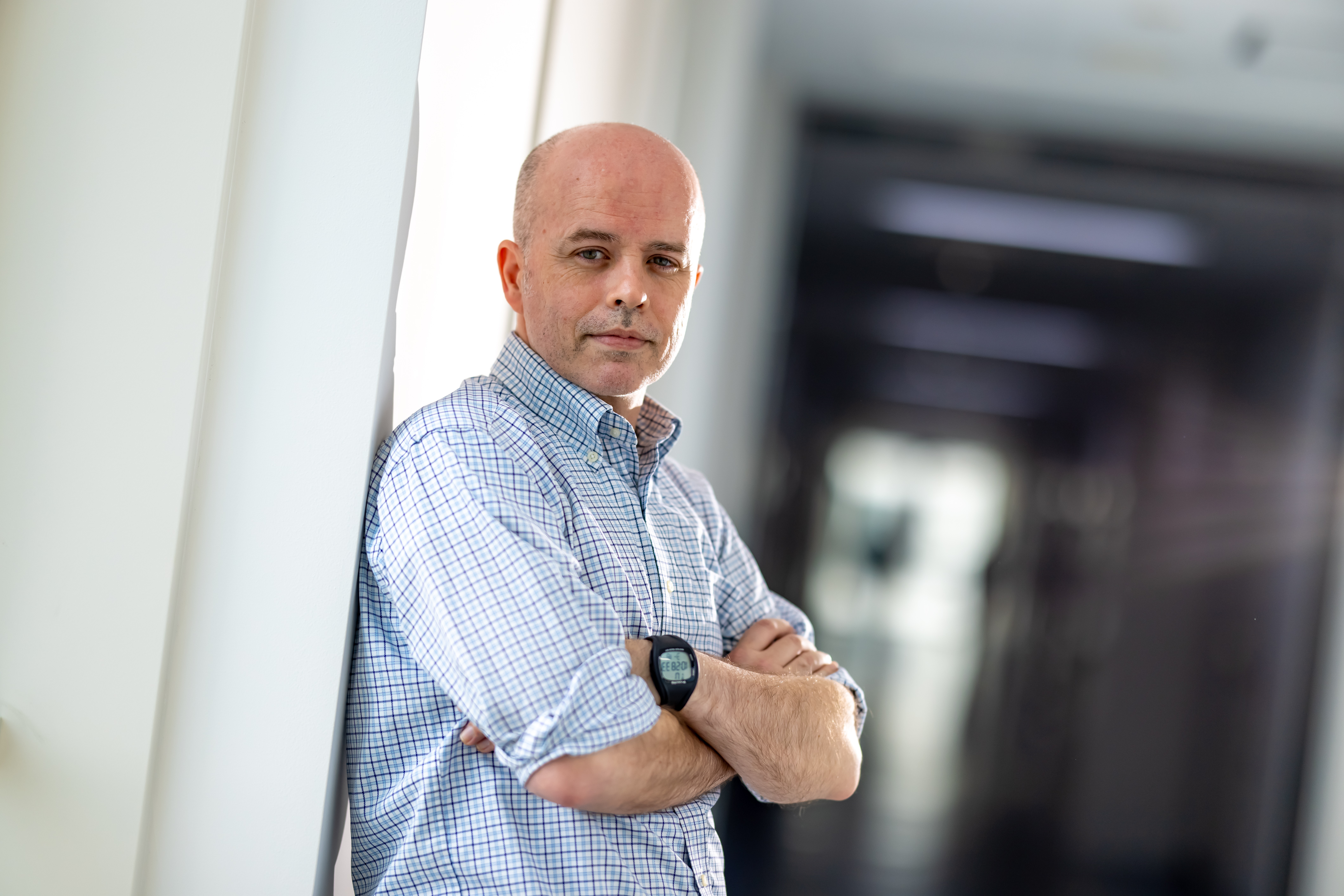
Dr. Nicholas I. Smith, Associate Professor, Biophotonics Laboratory, Immunology Frontier Research Center (IFReC)
"Physics meets biology in Biophotonics"
Born and raised in Australia, Dr. Nicholas I. Smith is the principal investigator of the Biophotonics Laboratory in the Immunology Frontier Research Center (IFReC). After completing an honors degree in physics and mathematics at the University of Sydney, he pursued a graduate degree at the same university before coming to Osaka University as a Ph.D. student in applied physics. Dr. Smith is trained in the field of optics and has been using his skills in spectroscopy and microscopy to understand biological phenomena. In particular, he is now using a combination of Raman spectroscopy, a technique commonly used in chemistry to identify molecules, with quantitative microscopy to address exciting problems in immunology.
Raman imaging: a combination of microscopy and Raman spectroscopy
Raman imaging is a label-free technique that Dr. Smith and his team have been using to retrieve measurable information of molecules that act as biomarkers of the morphological and molecular phenotypes of individual cells. Raman spectroscopy relies on inelastic scattering, which means that the energy of photons scattered from a molecule is not equivalent to that of the incident photons. The differences in energy depend on the molecule's vibrational modes, resulting in signature peaks of red-shifted electromagnetic spectrum that can be used to identify certain types of molecules. With the availability of highly sensitive detectors, it is now possible to perform high-speed laser scanning throughout the field of view of a microscope. The Raman spectrum of each pixel can then be analyzed to detect the main molecule constituents of various parts of a cell. Combined with computational analysis, this technique has opened up new ways to observe various states of a cell and identify new subtypes of immune cells.
It takes a village to produce good science
Dr. Smith has been collaborating with immunologists as well as physicists and chemists from Osaka University and other institutions to develop label-free and hybrid cell imaging techniques and utilize them to analyze various biological samples. One of the first projects that Dr. Smith worked on with IFReC scientists was the use of Raman imaging to observe hemozoin, a pigment formed from the digestion of red blood cells by blood parasites such as Plasmodium spp., the causative agent of malaria. From this work, Dr. Smith was invited to start an imaging lab at IFReC and has been working there with his team of scientists and graduate students ever since. In one of his current projects, his team utilizes Raman imaging and autofluorescence to observe the progress of immune cell activation. Dr. Smith appreciates the open research environment and administrative support at IFReC, and hopes that international researchers working in all parts of Osaka University enjoy the same or an even better experience.
Text: Clement Angkawidjaja/Edit: Christopher Bubb